Stereograms and the Standardization of Anatomical Observation: Arthur Thomson’s The Anatomy of the Human Eye, 1912
“One is hearing now, in this country at least, that in the near future we may look for a great revival of stereo photography, and if the rumor is well founded and turns out to be fulfilled prophecy, we may expect once more to find the stereoscope ‘on every drawing-room table,’ as of yore.” Thus wrote the photographic journalist Andrew Pringle in 1892, thirty years after the first, golden age of the stereoscope had passed. In the event, Pringle’s prediction would only be partially fulfilled, stereo photography never quite regaining the height of fashionability it once enjoyed in the drawing rooms of the middle classes and aristocracy. But revived it was, most significantly in the scientific rather than the domestic sphere.
The revival of stereo photography of the 1890s is less familiar to historians than the first flowering of the stereoscope during the late 1850s and 1860s. Nevertheless, like its predecessor, it was characterized by the publication of collections of topographical stereograms for the tourist and traveller, the release of treatises on the practical side of the subject aimed at amateurs, the manufacture of innovative equipment for both professional and consumer use, and much more. Scientific stereoscopy was not unknown during the mid Victorian period, as Warren de la Rue’s iconic stereograms of the moon testify. However, in one particular branch of science it was only at the turn of the century that its potential began to be fully explored: the science of medicine and anatomy.
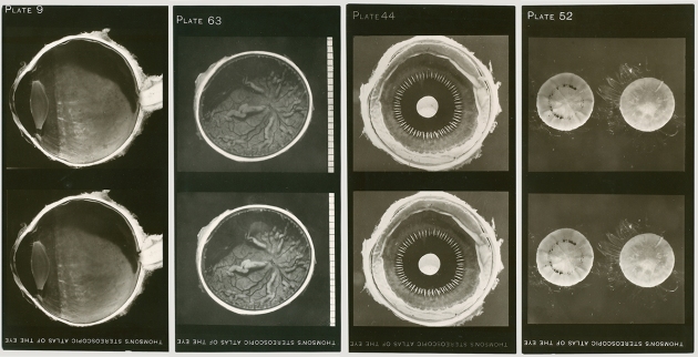
Among several innovative applications of stereo photography in medicine and anatomy in the period one in particular stands out due to the reflexive nature of its subject matter: Arthur Thomson’s The Anatomy of the Human Eye, as Illustrated by Enlarged Stereoscopic Photographs (Clarendon Press, Oxford, 1912). Also referred to as “Thomson’s Stereoscopic Atlas of the Eye”, the work consisted of sixty-seven “enlarged stereoscopic photographs” of human eyeballs in various states of dissection (see the gallery at the bottom of this article), together with a handbook of detailed descriptions and diagrammatic keys to the images.


The naissance of Thomson’s Atlas lay in his triangular interest in photography, art and anatomy. In March 1885 he was appointed the first Lecturer in Human Anatomy at Oxford, in November 1893 the first Professor. Simultaneously with his Oxford posts he also served as Lecturer in Anatomy at the National Art Training School in South Kensington (the Royal College of Art) and from December 1900 as Professor of Anatomy in the Royal Academy of Arts, at Burlington House on Piccadilly (a position he retained until 1934). His professional appointments reflected his lifelong interest in art, which also found expression in his love of watercolour painting and through his marriage to Mary, daughter of the portrait painter Norman Macbeth.
Sixteen years before compiling his Anatomy of the Human Eye, Thomson published a work that also combined photography, anatomy and drawing: A Handbook of Anatomy for Art Students (Clarendon Press, Oxford, 1896). The handbook, which proved popular and influential, differed from similar works in departing from an emphasis on the nomenclature and technical details of human anatomy in favour of the effect of anatomical structures on the surface forms of the body. To this end Thomson included “copious illustrations” in the volume, primarily employing photography for the purpose. For subjects he turned to “some of the better known athletes of this University” (who “for obvious reasons” were left unnamed), together with professional models (presumably the female sitters).


As well as stressing the importance of anatomy for artists, Thomson also advocated the cultivation of art for anatomists. In a letter to the British Medical Journal in 1896 attacking the General Medical Council’s allocation of marks in their examinations for proficiency in shorthand, he argued that “the use of drawing is an accomplishment of the highest value to medical men; for not only does it enable them to express in graphic form the appearance of an object, but its practice cultivates those powers of observation which are so necessary a feature in the training for our profession”. Photography he also viewed as much more valuable than shorthand: “take, for instance, the uses to which photography is now applied, as well might you expect a student to display a knowledge of these subjects, as tempt him, by the possible chance of scoring a few additional marks, to submit to an examination in photography.”
Prior to the publication of his Atlas Thomson explored the application of photography (via anatomy) to another of his academic interests: anthropology. In 1905 he published an article on “Composite Photographs of Early Egyptian Skulls”, explaining how he had used the technique in coming to the conclusions in his monograph, co-authored with the archaeologist David Randall-MacIver, on The Ancient Races of the Thebaid (Clarendon Press, Oxford, 1905). “It seems to me that after such a graphic demonstration [by composite photography] the question of the homogeneity of the early inhabitants of the Thebaid can no longer be maintained”, he argued in support of the view that the population of ancient Thebes (modern Luxor) had consisted of two separate races living side by side, the “negroid” and “non-negroid”.
Today Thomson is remembered primarily for his contribution to the establishment of medical teaching in Oxford, rather than for original research. In 1892 and 1893 he oversaw the design and construction of a new building for the Department of Human Anatomy, on the east side of the University Museum, which replaced a group of sheds occupied since 1885.


Thomson was keen to make his new accommodation available to colleagues in the University. When the British Medical Association met at Oxford in 1904, for example, the dissecting room was put at the disposal of Ernest William Ainley-Walker, tutor in physiology at University College, for a temporary “Pathological Museum”. Photographs featured prominently among the 1,000 exhibits displayed in the Museum, with a section dedicated to “photomicrographs and apparatus for photomicrography”, as well as, at an early date in its history, “colour photography in relation to medical, surgical, and pathological work”. According to the British Medical Journal, colour photography was “to the fore” during the meeting as a whole, the “demonstrations of the method”, by a “Mr Shepherd” (presumably Edward Sanger Shepherd), “even more instructive than the very beautiful examples of finished work.”
Stereo photography, in contrast to colour, was an old technology. However, during the 1900s it attracted renewed attention in the medical sciences, leading to the publication of several notable works illustrated partly or wholly by stereograms. In 1905, for example, David Waterston edited The Edinburgh Stereoscopic Atlas of Anatomy (T. C. and E. C. Jack, Edinburgh and London), which consisted of 250 stereograms, mounted on explanatory cards and housed in five boxes-cum-volumes. The Edinburgh Atlas was prepared “under the control” of the Department of Anatomy of the University of Edinburgh, where Thomson had preceded Waterston as Demonstrator in Anatomy, prior to moving to Oxford. In 1908 and 1909 Waterston’s work was followed by a more specialized study, the The Edinburgh Stereoscopic Atlas of Obstetrics (4 vols, T. C. and E. C. Jack), edited by G. F. Barbour Simpson and Edward Burnet. In 1910 Edinburgh’s monopoly on the genre was broken by Albert A. Gray, a Glasgow surgeon who authored The Ear and its Diseases (Baillière, Tindall and Cox, London). This was illustrated with thirty-seven stereoscopic illustrations Gray had taken himself and sold with a simple stereoscopic viewer included.


The immediate motivation for Thomson’s Anatomy of the Human Eye was not these works, but lectures on gross anatomy he was required to give to graduate students studying for the Diploma in Ophthalmology instituted at Oxford in 1910. The photographs he exhibited at these lectures were “so generally appreciated” by his students, he claimed, that he decided to publish them, in order to “make them accessible to those who would not otherwise have an opportunity of seeing them”. For Thomson’s students, who numbered 50 each term, his stereograms of the eye served as a “handy means of reference” when fresh specimens were unobtainable or permanent preparations inaccessible.
Photographic proxies were useful in part due to one of the drawbacks of medical study in Oxford, which according to the BMJ was the difficulty of finding raw material: “The supply of unclaimed bodies in Oxford itself is of the most limited kind, and guardians in surrounding districts do not show any particular readiness to further the cause of anatomical education; in some cases, indeed, they place difficulties in the way. An amendment of the Anatomy Act, taking the disposal of the unclaimed bodies of persons who die in workhouses out of the hands of guardians, seems to be required.”
The value of monoscopic photographs, in Thomson’s view, was largely self-evident and vested in photography’s unchallengeable objectivity. In his Handbook of Anatomy for Art Students, for example, he pointed out that the drawback of using photography from an artistic point of view was “counterbalanced by the truth of the resulting figures.” “The plates lay no claim to artistic excellence”, he confessed, rather “their value depends on their fidelity to nature.” Stereograms he justified on more practical grounds: in the flesh the minute structures of the eye could only be seen with the aid of a lens, which “usually involves their examination by monocular vision, and consequently entails a loss of that stereoscopic effect which is so necessary to determine their exact relations.”
For Thomson, the use of photographs in this way therefore served as a means of exercising greater control than was possible with real specimens over the observations his students made and how they interpreted what they saw. By complaining that the lack of material “in a sufficiently fresh condition” often prevented the detailed structure of the eye being visible in his classes, he betrayed his frustration with the difficulty of governing what his students witnessed in the dissecting room. In preparing the Atlas, he had been able to circumvent this difficulty by photographing most specimens within four hours of death. More significantly, he was also able to eliminate “doubtful examples” and avoid “faulty” specimens: a process that allowed him to manage the replication of results more closely than was possible in the flesh and therefore to further the standardization of his students’ observational experience.
By publishing his photographs Thomson extended this process beyond the local context of his teaching laboratory. His Atlas served to disseminate an authoritative anatomy of the eye constructed in Oxford, promoting as it did so his own privileged interpretation of how the organ should be described and interpreted. The provision of detailed texts and diagrammatic reductions in the accompanying booklet advanced the control he was able to exercise over these operations still further.
The strategy of publishing photographs of selected dissections is so ubiquitous in anatomical textbooks today that it is almost invisible. Nevertheless, it has a pedagogic history of its own, which ultimately extends to the prototypes that Thomson and other early advocates of anatomical photography produced. If the use of stereograms is now rare in scientific illustration as a whole, in the field of ophthalmology it remains relatively common, the eye appearing to offer a seemingly irresistible subject for such treatment. David G. Campbell and Peter A. Netland’s Stereo Atlas of Glaucoma (Mosby, St Louis, Missori, 1998), for example, is one of several reference works in the field illustrated throughout by stereograms, richly printed as colour half-tones.
Thomson’s Atlas depended not only on the stereoscopic revival of the 1890s and 1900s, but on access to facilities for the mass production of actual photographic prints, for which he could rely on the considerable institutional support and expertise offered by the University Press. As well as a classroom technology, his photography was also important institutionally, as a means by which he could advance the visibility and reputation of his own department, within and beyond the University.
For the modern viewer, Thomson’s stereograms are arguably as interesting from an artistic point of view as they are instructive in anatomy. Many have an abstract quality attractive to contemporary visual tastes (see, for example, plate 30 below), by virtue of which they can be incorporated within a retrospective historical genre of the “art of science”. Tempting as it is to read the cards as images of a pair of eyes, photographed side by side, in reality they each depict only a single eyeball, viewed from two perspectives. Binocular in one sense, they therefore remain resolutely monocular in another.
Gallery
The stereograms below can be seen in 3D without special apparatus. Click on an image to view in the gallery format; adjust the size of your browser window so each slide is approximately 17 cm wide; view from approximately 35 cm away; and allow your eyes to diverge (as if viewing a distant object) while maintaining focus on the screen (in the same way as viewing Magic Eye pictures). [21.09.2013. The gallery no longer displays properly, so I have switched the format to a slide show, but the stereoscopic effect should still be visible. GMH]
This slideshow requires JavaScript.
Further Reading
“Human Anatomy at Oxford”, British Medical Journal, vol. 2, no. 1712 (21 Oct 1893), pp. 902-903
Arthur Thomson, “The General Medical Council and Shorthand”, British Medical Journal, vol. 2, no. 1876 (12 Dec 1896), pp. 1747-1748
“The Pathological Museum”, British Medical Journal, vol. 2, no. 2274 (30 Jul 1904), pp. 257-258
Arthur Thomson, “Composite Photographs of Early Egyptian Skulls”, Man, vol. 5 (1905), pp. 65-67
“The Oxford Medical School”, British Medical Journal, vol. 1, no. 2373 (23 Jun 1906), pp. 1479-14
“Ancient Egyptian Race Controversy” (http://en.wikipedia.org/wiki/Ancient_Egyptian_race_controversy)
Roberta McGrath, Seeing Her Sex: Medical Archives and the Female Body (Manchester, 2002), ch. 5, “Other dimensions: The Edinburgh Stereoscopic Atlas of Obstetrics“
Cite this article: Giles Hudson, “Stereograms and the Standardization of Anatomical Observation: Arthur Thomson’s The Anatomy of the Human Eye, 1912″, Matters Photographical ([https://mattersphotographical.wordpress.com], 11 Sep 2013)

